
Control of heart contractions is mediated through a combination of autonomic innervation via the cardiac plexus as well as the conduction pathway through the heart muscle itself. To understand heart innervation, we must study the autonomic contribution to the cardiac plexus and how it influences the conduction system of the heart.
Cardiac Plexus
The cardiac plexus is divisible into a superficial cardiac plexus, between the aortic arch and the pulmonary artery, and a deep cardiac plexus, between the aortic arch and the tracheal bifurcation. The plexuses receive a combination of both sympathetic and parasympathetic fibers.
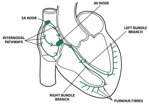
Autonomic innervation and conducting system of the heart.
Sympathetic Contribution
Preganglionic sympathetic fibers originate bilaterally in the lateral horns of the gray matter of the spinal cord between the T1 and the T5 spinal cord levels and enter the sympathetic chain via the white rami communicantes. After entering the sympathetic chain, fibers travel to the cardiac plexus via two possible routes:
- Preganglionic sympathetic fibers synapse in the superior parts of the thoracic sympathetic chain and send postganglionic sympathetic fibers directly from the sympathetic ganglia to the cardiac plexuses via thoracic cardiac nerves.
- The preganglionic sympathetic fibers ascend through the sympathetic chain and synapse in either the superior, middle, or inferior cervical ganglia before sending off postganglionic sympathetic fibers via cervical cardiac nerves to the cardiac plexuses.
 Referred pain. Both sympathetic and parasympathetic fibers carry visceral sensory fibers from the heart to the spinal cord and brain, respectively. However, the visceral sensory fibers within the cardiac branches from the cervical and superior five thoracic sympathetic ganglia are sensitive to ischemia. These sensory fibers mediate the visceral pain associated with angina pectoris and myocardial infarctions. Such myocardial ischemic pain is often referred to regions of the T1–T4 dermatomes simply because the visceral sensory fibers enter the spinal cord at the same levels of the segments for the superior four thoracic spinal nerves. The brain cannot differentiate between sensory input from the spinal nerves and that from the visceral nerves and thus refers ischemic pain to the same dermatome.
Parasympathetic Contribution
Preganglionic parasympathetic (vagal) fibers in the left and right vagus nerves originate in the medulla oblongata and descend through the neck and into the thorax to the cardiac plexuses. The synapse of vagal preganglionic and postganglionic parasympathetic fibers occurs either in the cardiac plexus or in the walls of the heart near the SA node of the right atrium. Therefore, the cardiac plexus serves as a conduit not only for parasympathetic preganglionic and postganglionic and visceral sensory fibers but also for sympathetic postganglionic fibers.
In summary, mixed nerves from the cardiac plexus supply the heart with sympathetic fibers, which increase the heart rate and the force of contraction and cause dilation of the coronary arteries, and parasympathetic fibers, which decrease the heart rate, reduce the force of contraction, and constrict the coronary arteries.
The autonomic branches from the cardiac plexus help to regulate the rate and force of heart contractions, through influencing the SA node and the AV node, as follows:
- SA node. The rhythm of the heart is normally controlled by the SA node, a group of automatically depolarizing specialized cardiac muscle cells located at the superior end of the crista terminalis, where the right atrium meets the superior vena cava. The SA node is considered the “pacemaker of the heart”. It is the point of origin of the electrical impulses that are propagated through the heart. It initiates the heart beat, which can be altered by autonomic nervous stimulation (sympathetic stimulation speeds it up, whereas vagal stimulation slows it down). The wave of depolarization sweeps down the walls of the atria, stimulating them to contract, and eventually reaches the AV node.
- The SA node automatically generates an electrical impulse 60-100 times per minute under normal conditions. These electrical impulses stimulate the atria to contract and then travel to the atrioventricular node (AV node), which is located in the inter-atrial septum.
- AV node. The AV node is located in the interatrial septum just superior to the opening of the coronary sinus. This node receives impulses from the SA node and passes them to the AV bundle (of His). Here the impulse is briefly slowed before continuing down the conduction pathway to the bundle of His.
- AV bundle (of His). The AV bundle begins at the AV node and descends through the fibrous skeleton of the heart before dividing into the left and right bundles (of His), corresponding to the left and right ventricles, respectively. This divergent pathway ensures that ventricular contraction begins in the region of the apex. Conduction ends near the aortic and pulmonic valves. Impulses also pass from the left and right bundle branches to the papillary muscles in the corresponding ventricles. In the right ventricle, the moderator band (septomarginal trabeculum) contains the right bundle branch. The bundle of His divides in the septum between the two ventricles into the left and right bundle branches, which are situated in the left and right ventricular muscle respectively.
- Purkinje fibers. Conduction then spreads out via specialized tissue within the venrticular walls known as Purkinje fibres.
The heart receives its arterial blood supply from the first branches of the ascending aorta, the right and left coronary arteries.The right and left coronary arteries arise at the aortic sinuses, the pockets formed by the right and left cusps of the aortic valve, respectively.
\
The right coronary artery (RCA) arises at the right aortic sinus and courses in the coronary (AV) groove between the right atrium and ventricle. At the level of the right auricular appendage, it gives off the SA nodal branch, which ascends to the junction of the SVC with the right atrium, where the SA node is located. As it reaches the inferior margin of the heart in the coronary groove, it will usually give off a right marginal branch that supplies the right ventricle along the inferior border. The RCA then curves around the inferior margin of the heart in the coronary groove onto the inferior and posterior surfaces of the heart, passing somewhat to the left toward the junction with the posterior interventricular groove, also called the crux of the heart. At the crux, the AV nodal branch passes deep into the interatrial septum to supply the AV node. The RCA divides into a larger posterior interventricular artery, which descends in the groove or sulcus of the same name. It passes toward but typically does not reach the apex of the heart. It supplies the right and left ventricles and posterior portions of the interventricular septum. A small branch continues to the left side of the heart to supply portions of the left atrium and ventricle and will anastomose with the circumflex branch of the left coronary artery (LCA) (see Figures 16-1 and 16-2).
The left main coronary artery divides quickly into the left anterior descending (LAD) and the left circumflex (LCx) arteries. The LAD artery courses anteriorly in the interventricular groove and gives rise to one, two, or three diagonal, and several septal, branches. The LAD itself terminates at the apex of the left ventricle. The diagonal branches of the LAD distribute blood to the anterolateral aspect of the heart. The septal branches of the LAD supply the interventricular septum, the bundle branches, and the Purkinje system. The other branch of the LMCA is the LCx artery, which courses along the left AV groove and gives rise to one to four obtuse marginal branches that supply the lateral wall of the left ventricle.
The anterior interventricular or left anterior descending (LAD) artery descends toward the apex of the heart in the anterior interventricular groove, where it curves around the apex onto the diaphragmatic surface of the heart to anastomose with the posterior (descending) interventricular branch of the RCA. The anterior interventricular artery supplies the anterior portion of the right and left ventricles and anterior two-thirds of the interventricular septum and therefore is the chief blood supply to the AV and the right and left bundles of the heart's conducting system. The other, smaller, branch of the LCA is the circumflex branch, which travels in the coronary groove toward the left margin of the heart, at which point it typically gives off a left marginal branch that supplies the left heart border portion of the left ventricle. The circumflex artery curves around the left heart border to anastomose with the RCA at the posterior aspects of the left atrium and ventricle. The pattern of arterial blood supply at this point is often described as a balanced blood supply because the RCA and LCA supply approximately equal amounts to the heart. In approximately 15 percent of the population, the LCA will supply a larger proportion than the RCA.
The majority of venous blood will enter the right atrium through the coronary sinus, which lies in the coronary groove on the posterior surface of the heart. Its internal opening is adjacent to the opening of the IVC. Great, middle, and small cardiac veins and several smaller named veins drain into the coronary sinus. A variable number of small anterior cardiac veins drain directly into the right atrium. The smallest cardiac veins drain small amounts of blood from the myocardial capillary plexus directly into the atria and ventricles.
The heart's pericardial sac receives its arterial blood supply primarily from the pericardiacophrenic artery (a branch of the internal thoracic artery) that accompanies the phrenic nerve. Small amounts of arterial blood are also provided by branches of the musculophrenic, superior phrenic, bronchial, and esophageal arteries. Pericardiacophrenic veins drain blood to the internal thoracic or brachiocephalic veins.

The anterior interventricular or left anterior descending (LAD) artery descends toward the apex of the heart in the anterior interventricular groove, where it curves around the apex onto the diaphragmatic surface of the heart to anastomose with the posterior (descending) interventricular branch of the RCA. The anterior interventricular artery supplies the anterior portion of the right and left ventricles and anterior two-thirds of the interventricular septum and therefore is the chief blood supply to the AV and the right and left bundles of the heart’s conducting system. The other smaller branch of the LCA is the circumflex branch, which travels in the coronary groove toward the left margin of the heart, at which point it typically gives off a left marginal branch that supplies the left heart border portion of the left ventricle. The circumflex artery curves around the left heart border to anastomose with the RCA at the posterior aspects of the left atrium and ventricle. The pattern of arterial blood supply at this point is often described as a balanced blood supply because the RCA and LCA supply approximately equal amounts to the heart. In approximately 15 percent of the population, the LCA will supply a larger proportion than the RCA.
The left coronary artery gives rise to the following two sizable branches:
- Left anterior descending artery. Also called the anterior interventricular artery and is often referred to by the acronym LAD. It supplies the anterior region of the left ventricle, including the anterolateral myocardium, apex, anterior interventricular septum, and the anterolateral papillary muscle.
- Left circumflex artery. Wraps around the left side to the posterior side of the heart. The circumflex artery supplies the posterolateral side of the left ventricle and gives off the left marginal branches which also supply the left ventricle.
Right Coronary Artery
The right coronary artery arises from the aorta, superior to the right cusp of the aortic valve. The right coronary artery travels along the right AV groove, between the root of the pulmonary trunk and the right auricle, and supplies the right atrium, right ventricle, the sinuatrial (SA) node, and the AV node. The right coronary artery gives rise to the following branches:
- Posterior descending artery. Supplies the inferior wall, posterior interventricular septum, and the posteromedial papillary muscle. In a few cases, the circumflex artery gives off the posterior descending artery.
- Right marginal artery. Supplies the right ventricular wall.
- SA nodal artery. Passes between the right atrium and the opening of the superior vena cava and supplies the SA node. In a few cases, the circumflex artery supplies the SA nodal artery.
Coronary Dominance
Coronary dominance is determined by which of the left and right coronary arteries gives rise to the posterior interventricular artery, which supplies the posterior interventricular semptum and part of the left ventricle. The dominant artery is usually the right coronary artery.
- In a balanced coronary circulation, the conduction system nodes of the heart (SA and AV nodes) are typically supplied by the RCA.
- In a balanced coronary circulation, the anastomoses between branches of the RCA and LCA occur at the posterior coronary and posterior interventricular grooves.
Questions
As a cardiologist, you are concerned about blockage of the artery to the SA node in a patient. This artery typically arises from which of the following?
RCA
Right marginal artery
Posterior interventricular artery
Anterior interventricular artery
Circumflex artery
In a balanced coronary artery pattern, the blood supply to the majority of the interventricular septum is derived from which of the following?
RCA
Internal mammary artery
Posterior interventricular artery
Anterior interventricular artery
Circumflex artery
The correct answer is D. You answered D.
D. Usually, the anterior two-thirds of the interventricular septum is supplied by the AV artery, and the right and left bundle branches of the conduction system are generally supplied by the anterior interventricular artery.
As a cardiologist, you are concerned about blockage of the artery to the AV node in a patient. This artery typically arises from which of the following?
RCA
Right marginal artery
Posterior interventricular artery
Anterior interventricular artery
Circumflex artery
The correct answer is A. You answered D.
A. The AV node is also supplied by the RCA.
A 56-year-old man is complaining of chest pain that radiates to the jaw and left arm. A thallium stress test shows decreased perfusion to the heart that overlies the diaphragm. Which of the following coronary arteries is most likely to be blocked?
Left anterior descending
Right coronary
Left main artery
Left circumflex
The correct answer is B. You answered B.
B. The inferior portion of the heart is supplied by the right coronary artery.
Study Questrions
A 62-year-old man is brought to the emergency department after experiencing a myocardial infarction. His heart rate is 40 beats/min. Further examination reveals an occlusion of the patient's right coronary artery. Which of the following structures is most likely affected by this blockage?
AV node
Bundle of His
Mitral valve
Tricuspid valve
The correct answer is A. You answered A.
A myocardial infarction in the inferior wall involving the right coronary artery may affect the AV node, resulting in bradycardia.
Content 2
Content 3
The majority of venous blood will enter the right atrium through the coronary sinus, which lies in the coronary groove on the posterior surface of the heart. The internal opening of the coronary sinus is adjacent to the opening of the IVC. Partly, small veins empty into the right atrium. The coronary sinus, runs from left to right in the posterior part of the coronary sulcus. The coronary sinus receives the great cardiac vein at its left end and the middle cardiac vein and small cardiac veins art its right end. The left posterior ventricular vein and left margical vein also open into the coronary sinus.

(
Great, middle, and small cardiac veins and several smaller named veins drain into the coronary sinus. A variable number of small anterior cardiac veins drain directly into the right atrium. The smallest cardiac veins drain small amounts of blood from the myocardial capillary plexus directly into the atria and ventricles.
The cardiac veins and associated tributaries are the major veins of the coronary circulation and run parallel to the arteries. They drain blood from the heart wall. The cardiac veins are as follows:
- The coronary sinus is the largest vein draining the heart muscle and lies in the coronary sulcus. The coronary sinus collects most of the venous return from the great, middle, and small cardiac veins and returns the venous blood to the right atrium. The coronary sinus opening in the right atrium is superior to the septal leaflet of the tricuspid valve.
- Great cardiac vein. Begins at the apex of the heart and ascends in the anterior interventricular groove, parallel to the left anterior descending artery, and drains into the coronary sinus.
- Middle cardiac vein. Begins at the apex of the heart and ascends in the posterior interventricular sulcus, parallel to the posterior interventricular artery, and drains into the coronary sinus.
- Small cardiac vein. Courses along the acute margin of the heart, along with the marginal artery, and then courses posteriorly into the coronary sinus.
- The anterior cardiac veins drain the anterior portion of the right ventricle, cross the coronary groove, and empty directly into the right atrium. Anterior cardiac veins do not drain into the coronary sinus.
The normal pericardium is a double-layered sac.
The visceral pericardium adheres firmly to the epicardium, reflects over the origin of the great vessels,is a serous membrane that is separated by a small quantity (15–50 mL) of fluid, an ultrafiltrate of plasma, from the fibrous parietal pericardium.
The normal pericardium, by exerting a restraining force, prevents sudden dilation of the cardiac chambers, especially the right atrium and ventricle, during exercise and with hypervolemia.
It also restricts the anatomic position of the heart, and probably retards the spread of infections from the lungs and pleural cavities to the heart.
Nevertheless, total absence of the pericardium, either congenital or after surgery, does not produce obvious clinical disease.
In partial left pericardial defects, the main pulmonary artery and left atrium may bulge through the defect; very rarely, herniation and subsequent strangulation of the left atrium may cause sudden death.

Computed tomography scan shows the normal pericardium as a thin, curvilinear line (open arrows). The increased thickening over the anterior surface of the heart (solid arrows) is probably an artifact from transmitted right ventricular (RV) pulsations.
The pericardial space is enclosed between these two serosal layers and normally contains up to 50 mL of a plasma ultrafiltrate, the pericardial fluid. Pericardial reflections around the great vessels tether the pericardium superiorly and result in the formation of two potential spaces: the oblique and transverse sinuses. Superior and inferior pericardiosternal and diaphragmatic ligaments limit displacement of the pericardium and its contents within the chest and neutralize the effects of respiration and change of body position. The phrenic nerves are embedded in the parietal pericardium and, for this reason, are vulnerable to injury during pericardial resection.
Which of the following vessels is responsible for transporting oxygenated blood from the lungs to the heart?
The correct answer is E. You answered E.
Oxygenated blood is transported from the lungs to the left atrium via the pulmonary veins.
Reference: http://www.digital-world-medical-school.net/01.%20Medical%20School/1.%201st/09.%20Regional%20Gross%20Anatomy/03.%20Thorax/03.%20Heart/Heart.html
A contrast study of the pulmonary vessels will most likely reveal several pulmonary veins entering the left atrium. How many pulmonary veins entering the left atrium will most likely be seen?
Two
Three
Four
Five
Six
The correct answer is C.
At the wall of the left atrium four pulmonary veins deliver oxygenated blood into the left atrium.
Reference: http://www.digital-world-medical-school.net/01.%20Medical%20School/1.%201st/09.%20Regional%20Gross%20Anatomy/03.%20Thorax/03.%20Heart/Heart.html/#Chambers
Reference:
During the autopsy of a trauma victim, the pathologist noted a tear at the junction of the superior vena cava and the right atrium. Which of the following structures would most likely have been damaged by the tear?
Atrioventricular (AV) bundle
AV node
Left bundle branch
Right bundle branch
Sinuatrial (SA) node
The correct answer is E. You answered B.
The SA node, or pacemaker, lies within the right atrial wall, where the right atrium is joined by the superior vena cava.
Reference : http://www.digital-world-medical-school.net/01.%20Medical%20School/1.%201st/09.%20Regional%20Gross%20Anatomy/03.%20Thorax/03.%20Heart/Heart.html
In a healthy person, blood from the pulmonary trunk will flow next into which of the following structures?
The correct answer is D.
Deoxygenated blood from the right ventricle is pumped into the pulmonary trunk, which bifurcates into the right and left pulmonary arteries before coursing to the lungs.